Animals were of prime importance in the classical world: agriculture in the Roman Empire depended on domesticated oxen (L., bos) to plow the fields, crowds of thousands cheered on the races at the Circus Maximus, as drivers atop their chariots urged on the fastest horses (L., equus) in the land, and sacrifices of sheep (L., ovis), and pigs (L., porcus) were necessary to please the gods. So too were animals important to ancient scientists and physicians. It was the dissection of and the experiments on animals that formed the basis for early understanding of human anatomy and physiology. In addition, there are countless anatomic terms whose etymologies derive from the Greek and Latin names of animals.

Cauda equina
Origin: Latin, cauda (tail) + equus (horse)
Horse’s tail

cauda equina

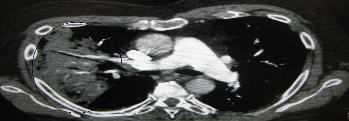
Sitting within and protected by the vertebral column, the spinal cord consists of nerve cells and their extensive myelinated tracts that carry motor, sensory, and autonomic data to and from the brain. One would think that the spinal cord extends all the way to the bottom of the vertebral column, but in fact it stops looking like a cord at the level of the L2 vertebra in adults. At that point, it gives rise to the so-called cauda equina where individual nerves arising from the lower part of the spinal cord descend down the vertebral column until they exit through the proper foramina. It’s no wonder then why lumbar punctures must be done below L2: the individual nerves of the cauda equina can easily accommodate the lumbar puncture needle unlike the large-diameter spinal cord. So the next time you consent a patient for an LP, be sure to ask yea or neigh.

Coccyx
Origin: Greek, kokkux (cuckoo)

sacrum

The vertebral column consists of seven cervical, twelve thoracic, and five lumbar vertebrae. The primary role of these bones is to protect the spinal cord. However, their articulations allow flexion, extension, lateral motion, and rotation of the back, explaining their derivation from vertere (L., to turn). There are two more bones that lie below L5. The sacrum, which the Romans called the os sacrum (L., sacred bone) and which the Greeks named the hieron osteon (Gk., sacred bone), sits within the pelvis and allows the sacral nerves of the cauda equina to exit through its foramina. It’s not clear why this bone was considered “sacred.” One thought is that its sacredness derives from its location in the pelvis and therefore an association with reproduction. Another theory suggests that the ancients used this bone during sacrificial ceremonies. Finally, below the sacrum sits the coccyx, the small bony remnant of a vestigial tail. The ancient anatomists who named this bone thought that, when viewed from its side, it bears resemblance to a cuckoo’s beak.

Tragus
Origin: Greek, tragos (goat)

traigus

The tragus is that small piece of cartilaginous tissue situated next to the opening of the ear canal. And for some strange reason, it’s derived from the Greek word for goat. Perhaps some Greek anatomist had particularly hairy ears and thought it resembled the hair on a goat’s chin. Whatever the reason, the association with goats definitely stuck: the medical term describing hair on the ears is barbula hirci, literally meaning “little beard of the goat” [L., barbus (beard) + hircus (goat)]. No kidding.

Concha
Origin: Greek, konkhe (mussel, shell)

concha

Within the nose lie the superior, middle, and inferior nasal conchae. These long shelves of bone on the lateral aspects of the nasal cavity are covered with mucosa and supplied by a rich network of blood vessels. Their most important function is to warm and humidify the air as it passes from the nasal cavity to the lungs. If I look really hard, I can make myself believe they look like little mussels. Astute readers will notice that concha also appears in the labeled anatomy of the outer ear: concha also describes the fossa next to the opening to the ear canal.

Pes Anserinus
Origin: Latin, pes (foot) + anser (goose)
Goose’s foot

pes anserinus

Three muscles of the leg, the gracilis [L., gracilis (slender)], sartorius [L., sartor (tailor)], and semitendinosus [L., semi (half) + tendere (to stretch)], give rise to tendons that travel in parallel before attaching to the same location on the tibia. The ancient anatomists who saw this arrangement must have been reminded of the tripartite webbed feet of the goose and named it as such. There is a clinical condition called “pes anserinus pain syndrome,” also known as “pes anserine bursitis,” that describes pain around these tendons on the medial side of the knee. Interestingly enough, pes anserinus is not only a structure in the leg: it also is another name for the parotid plexus, where the facial nerve branches after exiting the stylomastoid foramen.

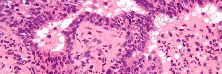
Recurrent Laryngeal Nerve

galen dissection

Galen demonstrating the effects of the recurrent laryngeal nerve during the dissection of a pig

The recurrent laryngeal nerves are branches of the vagus nerves that control nearly all of the intrinsic muscles of the voice box. Therefore, injury to one or both may cause hoarseness or even a complete inability to phonate. The etymology is nothing to write home about, but the story of their discovery definitely is. The famed Greek physician Galen, while conducting an experiment on a live, strapped-down pig, accidentally cut both laryngeal nerves; the pig continued to struggle but completely stopped squealing. He ultimately traced out the path of the nerves and confirmed that they originated from the vagus nerves. He noted that “there is a hairlike pair [of nerves] in the muscles of the larynx on both left and right, which if ligated or cut render the animal speechless without damaging either its life or functional activity.” Galen held a public demonstration of his experiment in Rome that was well-attended by distinguished politicians and scholars of the time. Those readers interested in learning more can read the article “Galen and the Squealing Pig,” linked here.

Thanks for taking a trip to the zoo with me. I’m off to see the capybara exhibit.

P.S.: Can you think of the English adjectives derived from the Latin words mentioned in the first paragraph? As a bonus, what about the adjective that describe wolves? Fish? Ducks?

Pneumonia is one of those medical conditions that is constantly thrown around without being rigorously defined—and as we all know by now, rigor is absolutely paramount in my view of academic medicine and medical etymology. Let’s start by looking at the word from an etymological standpoint:

Pneumonia
Origin: Greek, pneumon (lung) + –ia (condition of)
Condition of the lung

Well, that certainly wasn’t very helpful—at least not at first glance. There can be many conditions of the lung that are called “pneumonia” and the classic case of a lung infection is merely one of them. Compare pneumonia to pneumonitis:

Pneumonitis
Origin: Greek, pneumon (lung) + -itis (inflammation of)
Inflammation of the lung

There are many inflammatory conditions of the lung, but only a few of them are called “pneumonitis” because pneumonitis is considered to be inflammation of the lung tissue itself (the lung parenchyma). There are some who then consider pneumonia to be inflammation of the air sacs in the lungs (the alveoli). This makes sense for the most part; the bacteria, viruses, fungi, and parasites that can cause pneumonia would cause alveolar inflammation.

lung diagram

To put things in perspective, when somebody is admitted to the hospital for pneumonia, the classic picture is someone who acquires a bug, and said bug colonizes the alveolar space. The resulting “mass” that forms (usually in one lobe of the lung) is called a consolidation:

Consolidation
Origin: Latin, con- (with) + solidus (solid)
With solid, i.e., things that aggregate to form a “solid”

For these “classic” pneumonias, we can call them community-acquired pneumonia (CAP), hospital-acquired pneumonia (HAP), and the like. Some bugs associated with the former are things like Streptococcus pneumonia and Pseudomonas aeruginosa, and some bugs associated with the latter are things like Staphylococcus aureus and Haemophilus influenzae. (Stay posted for microbiological etymology!)

But it turns out that this isn’t the whole picture. Pneumonia is indeed as general as its etymology suggests, for there can be inflammatory and fibrotic conditions of the lung—and both classes can fall under the term “pneumonia.” (Generally, inflammation is caused by the infiltration of white blood cells and fibrosis is caused by the production of excess connective tissue, e.g., collagen.) Thus, as a counterexample to the definition that pneumonia is simply the inflammation of the alveoli, we have something called usual interstitial pneumonia (UIP). It is a form of interstitial lung disease in which the tissue between the alveoli is fibrosed with collagen.

uip ctThe classic “honeycomb lung” appearance of UIP on CT scan

To end this post, I posit my generalized definition of pneumonia: pneumonia is a principally inflammatory or fibrotic condition of the lung. By formalizing this definition of pneumonia, I am faithful to the etymology and make pneumonitis a subset of pneumonia. Note that I use the word “principally” in front of inflammatory—the reason why is that cancers of the lung can cause inflammation, but cancers are not considered to be pneumonias by any definition or practice.

In another post I will discuss the classic methods of diagnosing “normal” (generally bacterial) pneumonia and the etymologies of these techniques. Here is a small teaser: one of the terms involves the Greek word for goat! Can you guess which one? Leave a comment below.

Playing the singular/plural game is always fun (at least to me), so here is a short aside with a few of my favorites that relate to the topics I lectured on these past few weeks:

  1. Apparatus → apparatus
    Origin: Latin, ap- (toward) + parare (to set)
    A piece of equipment, i.e., something that “sets you toward something”
  1. Foramen → foramina
    Origin: Latin, forare (to bore)
    An anatomical hole or opening
  1. Fossa → fossae
    Origin: Latin, fodere (to dig)
    An anatomical ditch, with only one open end
  1. Octopus → octopodes
    Origin: Greek, octo– (eight) + podos (foot)
    The animal with eight feet

Can you think of the plurals for these words? Leave a comment below!

  1. Plexus
  2. Isthmus
  3. Duodenum
  4. Platypus
  5. Rhinoceros (!)

There are a number of ways of diagnosing disease: tissue biopsy, symptom matching, and of course, clinical signs. People often use the latter two terms interchangeably, especially since they are often paired together via the phrase “signs and symptoms.” But rigorously, these two words refer to two different things. Perhaps their etymologies will help differentiate them:

Sign
Origin: Latin, signum (sign/image)
Something the clinician notes in during a clinical encounter

Symptom
Origin: Greek, sym- (together) + piptein (to fall)
Something a patient reports to the clinician; something that “falls together” with something else

In other words, symptoms are subjective experiences while signs are objective experiences. For example, if I tell my doctor I have a fever, that is considered a symptom, but if he or she were to measure my temperature and find it elevated, it would be a sign. Another good example: pain is a symptom, since the perception of pain is unique to each patient. Tenderness, however, is a sign, since it is elicited by a clinician, e.g., applying pressure to the abdomen and eliciting an “ouch!”

stigmata rev

St. Francis Receives the Stigmata, by Antoni Viladomat (1678-1755)

There is another pair of terms that are often used together that is also often conflated. These words are stigma (plural stigmata) and sequela (plural sequelae). A stigma is a readily-visible (cutaneous or otherwise) or discernable sign that is characteristic of some disease. A classic stigma of liver disease is jaundice.A sequela, on the other hand, is an abnormal state or complication that results from a previous disease. A classic sequela of diabetes is diabetic nephrophaty. The etymologies of these words are relatively straight-forward:

Stigma
Origin: Greek, stizein (to mark/tattoo)

Sequela
Origin: Latin, sequi (I follow)

Interestingly enough, the word “stigmata” is also used in theology to refer to the bodily marks or sensations of pain associated with the crucifixion [Origin: Latin, crux (cross) + figere (to fix/bind)] of Jesus. Those who bear these stigmata are called “stigmatics” or “stigmatists,” and a common motif in classical painting is St. Francis of Assisi’s reception of the stigmata (see above). St. Francis supposedly received the stigmata of Christ for his great piety during the Feast of the Exaltation of the Cross after a forty-day fast.

Regarding jaundice, a stigma of liver disease, we know it to be the yellowing of the skin due to increased levels of bilirubin. Jaundice can, of course, be caused by a number of other conditions, e.g., deficiencies in certain enzymes (cf. Gilbert syndrome). There is one part of the body that is not said to be jaundiced when yellow; that honour falls on the eyes. When the eyes turn yellow due to, say, liver disease, we call it scleral icterus.

icterus rev

Jaundice
Origin: Latin, galbinus (yellow/yellow-green)

Icterus
Origin: Greek, ikteros
A bird said to cure jaundice when spotted

Hence we call it scleral icterus because it is with our eyes that we see the ikteros and become cured of our jaundice!

Meningitis (plural meningitides) is a nasty condition. From the first post, we already know that meningitis is defined as the inflammation of the meninges. The meninges (singular meninx) are the protective coverings of the brain, and there are three layers, listed outermost to innermost:

  1. Dura mater (Latin, “hard mother,” since it is the thickest layer)
  2. Arachnoid mater (Neo-Latin, “spider-like mother,” since it resembles a spider web)
  3. Pia mater (Latin, “pious mother,” erroneous translation from Arabic umm raqiqah for “tender mother”)

The dura mater is also known as the pachymeninx and the arachnoid mater + pia mater are also known as the leptomeninges.

meninges

Pachymeninx
Origin: Greek, pachy (thick) + meninx (membrane)
Thick membrane, i.e., the dura mater

Leptomeninges
Origin: Greek, leptos (thin) + meninges (membranes)
The thin membranes, i.e., the arachnoid mater and the pia mater

Meningitis can be caused by bacteria, viruses, autoimmune phenomena, or trauma. If the inflammation is caused by bacteria, it is said to be (quite logically) a bacterial meningitis, but if it is caused by the latter three, it is said to be an aseptic meningitis.

Aseptic
Origin: Greek, a- (without) + sepein (to cause decay)

Though the other aetiologies “cause decay,” septic is reserved for bacteria in the medical lexicon. It is important to note that the aseptic meningitides, though wretched, do not typically cause mortality. The bacterial meningitides, however, must be identified and treated swiftly, for they have a relatively high mortality. It is for this reason that if a case of bacterial meningitis is suspected, antibiotics are indicated for immediate use, whether or not the patient truly has a bacterial meningitis. Neutrophils in the cerebrospinal fluid (CSF) is a classic diagnostic marker of bacterial meningitis:

csf pleo rev

There are numerous signs that can hint towards a patient suffering from meningitis, but two of the classic ones are nuchal rigidity (neck stiffness) and photophobia.

Nuchal
Origin: Arabic, nukah (spine)
Used in English as an adjective for the neck

Photophobia
Origin: Greek, photos (light) + phobos (fear) + -ia (condition of)
Condition of the fear of light

When you think “vessel” and “neck,” what words come to mind? For me, the first two words I think of are carotid and jugular. I’ve heard these words used in the vernacular every so often, especially the latter. If someone wants to convey a sentiment of “going for the kill” or delivering a coup de grace, that person can say he or she is “going for the jugular.” Let’s start with this one:

Jugular
Origin: Latin, jugulum (throat)
Named for the fact the jugular veins are near the throat

jugular

There are two jugular veins, one external and one internal on each side of the body. The internal jugular is covered by the sternocleidomastodeus muscle, while the external one is readily visible. These two veins are extremely important because they drain the deoxygenated blood from the head back to the superior vena cava.

What about the other vessel(s) of interest? There is a common carotid artery on both sides of the body, and this artery branches into the internal and external carotid arteries. The former supplies blood to the brain, while the latter supplies blood to the face and head. The etymology is rather interesting:

Carotid
Origin: Greek, karos (stupor)
So called from Galen’s observation that their compression causes stupor/somnolence

carotid rev

If one were to bilaterally massage both carotid sinuses (the point at which the common carotid bifurcates), the baroreceptors of the sinus are fooled into thinking there is a higher-than-normal blood pressure. To compensate, the blood pressure decreases, resulting in a sensation of stupor. Interestingly enough, there is a therapeutic use for this maneuver: the alleviation of supraventricular tachycardia:

Supraventricular tachycardia
Origin: Latin, supra– (above) + ventricle (chamber of the heart; translates to belly, for the ventricles comprise the “belly” of the heart)
Origin: Greek, tachys (swift) + kardia (heart)
A fast pulse due to improper electrical activity above the level of the ventricles

What other afflictions could these vessels suffer from? One thing that can go wrong with the carotid artery is carotid artery stenosis:

Stenosis
Origin: Greek, stenos (narrow/close) + -osis
Condition of narrowness, taken to mean a narrowing

Carotid artery stenosis is oftentimes caused by atherosclerosis, or the reduction of the interior arterial (luminal) diameter due to the accumulation of gunk. This gunk is a lovely mixture of dead cells, cholesterol, and fats.

Atherosclerosis
Origin: Greek, athres (gruel) + sklerein (to harden) + –osis
Condition of the hardening of gruel

Sounds delicious!

What on earth is pyelonephritis? Let’s understand what it actually is by taking a look at its etymological breakdown.

Pyelonephritis
Origin: Greek, pyelos (basin, taken to mean pelvis) + nephros (kidney) + –itis (inflammation of)
Inflammation of the basin and the kidney

At first, this seems cryptic. What does basin refer to, and what exactly does kidney refer to? Starting with the root pyelo-, it denotes “pelvis,” but its base etymology comes from the Greek pyelos (basin). There is in fact such a thing as pyelitis, which would be inflammation localized to the renal pelvis. As for the nephr- root, it of course means kidney, but to be more precise, it refers to the kidney parenchyma. Nephritis on its own denotes the inflammation of the kidney’s functional tissue.

kidney anatomy rev

At the end of the day, pyelonephritis is the joint inflammation of the renal pelvis and the renal parenchyma. A vast majority of the cases are caused by a bacterial agent such as E. coli., and as such, they proliferate in a large portion of the kidney instead of being localized to only the pelvis or the parenchyma.

There are three types of pyelonephritis: acute, chronic, and xanthogranulomatous. Let’s look at the last one:

Xanthogranulomatous
Origin [1]: Greek, xanthos (yellow)
Origin [2]: Latin, granum (grain) + –ula (diminutive) + –oma (mass/proliferation/tumor)
Forming yellow granulomas

Indeed, under the microscope, one would find granulomas—collections of macrophages that try to wall off infections they cannot eradicate. On gross appearance, these granulomas appear yellow because they are macrophages that have been filled to the brim with lipids.

xantho rev

Diabetes is a household name in this day and age. More often than not, people are concerned about their sugar intake in order to prevent diabetes. A quick Google Scholar search will reveal that a high sugar intake does not increase the risk of developing diabetes. But, of course, a chronically-high concentration of glucose in the blood will lead to a variety of problems down the line. An age-old clinical observation of diabetes is the tendency to urinate often, termed polyuria [Greek: poly (many) + ourein (to urinate)]. This is in fact where the word diabetes comes from:

Diabetes
Origin: Greek, dia- (through) + banein (to pass)
To pass through, siphon

What about the two words appended to the end of “diabetes”? They are the following:

Mellitus
Origin: Latin, mel (sweet thing)
Something sweet, honey

Insipidus
Origin: Latin, in- (without) + sapere (to have taste)
Lacking taste, in this case sweetness

Diabetes mellitus (DM) has two types, Type 1 and Type 2. The former is usually ascribed to insulin insufficiency, whereas the latter is usually ascribed to insulin resistance. It is given the name “mellitus” because of glucose in the urine, or glucosuria. The large amounts of glucose in the blood finds its way to the urine, and as a result, makes the urine—the entity being passed through—sweet. The polyuria is caused by the osmotic effect of water “following” the glucose. The quick and easy way that physicians would diagnose diabetes mellitus was to simply taste the urine of a patient suspected to have this condition.

vasopressin rev

On the contrary, diabetes insipidus (DI) is caused by a lack of water control and is not related to diabetes mellitus. This “lack of water control” involves problems with anti-diuretic hormone (ADH) production or ADH insensitivity. The former is termed central DI, since it originates in the central nervous system, and the latter is termed nephrogenic [Greek: nephros (kidney) + -genes (born from)] DI. This lack of mechanistic control stimulates profound thirst to replace the massive amounts of water lost in the urine since the water is not being reclaimed by the distal convoluted tubule and collecting duct of the nephron. However, blood glucose is not affected by this condition, there is no glucosuria, and in turn there is no sweetness in the urinehence the urine is without taste, or “insipid.”

In colloquial language, we often append the suffix ­-itis to a word to denote a disease or condition of said word. It is in effect an indication of an unhealthy amount of something either abstract or tangible. If someone is being lazy, we can say that he or she has come down with a case of lazyitis. Too much time in front of a computer can afflict you with a nasty case of computeritis. At its roots, –itis means “pertaining to” in Greek, so in some sense, this colloquial use of the suffix is the most faithful to its base etymology.

In more technical language, –itis is used as a medical suffix, and it simply denotes “inflammation of.” I can’t tell you how many times I have heard people use ­–itis to immediately denote “infection of.” Let’s list some examples.

Appendicitis
Origin: Neo-Latin, appendix (something appended, in this case to the colon) + -itis (inflammation of)
Inflammation of the appendix

Bursitis
Origin: Greek, byrsa (hide/skin) + ­-itis
Inflammation of the bursa (a pouch or sack that facilitates motion, e.g., suprapatellar bursa)

Cholecystitis
Origin: Greek, chole (bile) + kystis (bag) + -itis
Inflammation of the gall bladder (which stores bile)

Yes, many times, these conditions come about from infection by a bug that isn’t supposed to be there. But there are many other causes of inflammation, such as:

  1. Trauma
  2. Autoimmune reaction
  3. Drugs
  4. Burns
  5. Radiation

So the next time someone mentions an ­–itis, always remember that the inflammation of the target organ can be caused by something other than infection. If you wish to convey the fact that an inflammation of the appendix is caused by an infection, you can prepend the word with “infectious,” to make it an infectious appendicitis. Or if you are more certain of the cause of this infection, you can use a bacterial/viral/fungal modifier, for a bacterial appendicitis. For a non-infectious cause, you could say the cause for an inflammation of the liver is a drug-induced hepatitis.

Finally, if someone mentions a type of ­­itis that you are not familiar with, use the word itself to help you out. I remember hearing the term bronchiolitis last year when learning about COPD (chronic obstructive pulmonary disease). I didn’t know what bronchiolitis was until I stopped for a second to think about it: the inflammation of the bronchiole(s). Contrast this with bronchitis, which is the inflammation of a main stem bronchus.

lung anatomy rev

So, given this, let me know in the comments what these terms are and what their most common causes are:

  1. Cholangitis
  2. Colitis
  3. Pneumonitis
  4. Endocarditis
  5. Vasculitis
  6. Phlebitis
  7. Gastroenteritis