In the medical field, there is a consistent misuse of the words hypertrophy and hyperplasia. It’s not quite as egregious as the misuse of the suffix “-itis,” but it is still saddening—at least to me. An etymological breakdown can help us differentiate between the two terms:

Hypertrophy
Origin: Greek, hyper (over/above) + trephein (to feed)
To overfeed ; taken to mean an increase in cell size

Hyperplasia
Origin: Greek, hyper (over/above) + plassein (to mould)
To mould over; taken to mean an increase in cell number

These are the definitions on the cellular level, but it turns out that some pathologists also use the term hypertrophy to denote a gross increase in organ size, hence the confusion. For example, when a man’s prostate becomes enlarged, it is due to hyperplasia (an increase in cell number). The cellular term for this phenomenon is nodular hyperplasia. On an autopsy report, however, a pathologist would report it as prostatic hypertrophy because he or she can see the increase in size through the naked eye. Hyperplasia is a normal physiological response and it is not pathologic. A good example is the hyperplasia of the uterine lining (the endometrium) in the menstrual cycle.

hyperplasia comparison rev

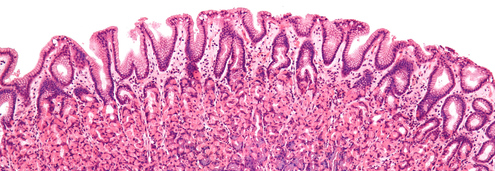
Note, however, that the story with hyperplasia is a bit more nuanced than that. In the case of the endometrium [Origin: Greek, endo- (in) + metron (womb)], physiologic hyperplasia is termed “simple hyperplasia without atypia.” The term simple vs. complex denotes that the cells either have or have not proliferated to the point of interfering with the normal architecture of the endometrium. The modifier of “atypia” denotes a structural abnormality of the cells; thus, atypical cells can be considered to be dysplastic.

This brings us to another pair of -plasias that is often confusing to people: neoplasia vs. dysplasia. The difference between these two terms is a bit more subtle, but again, some etymology may be of use to us.

Neoplasia
Origin: Greek, neo– (new) + plassein (to mould)
A new moulding; taken to mean an abnormal proliferation of cells (either benign or malignant)

Dysplasia
Origin: Greek, dys– (ill) + plassein (to mould)
An ill molding; taken to mean an increased number of immature cells (compared to regular cells) and greater variability between cells

On a functional basis, dysplasia may progress to neoplasia, but dysplasia by itself doesn’t denote an abnormal growth or increase in cell number. Indeed, dysplasia results in an increased ratio of immature to mature cells and variability because it has acquired just enough mutations to appear odd—hence, depending on the situation, its progression to neoplasia. One might then ask: “What about carcinoma in situ (CIS)?” Well, dear reader, CIS is a subset of neoplasia (abnormal proliferation of cells), but one that has not invaded through the basement membrane:

cis rev

There are other types of “–plasias,” such as metaplasia & desmoplasia. What do you think these terms denote and where do they come from? Let me know in the comments section!

If you want to do well on Step 1 of the USMLE, you will need to be able to answer two-step reasoning questions like the following:

A 38 year old morbidly obese woman presents to neurology clinic complaining of difficulty reading in bed at night. She has noticed that she is unable to keep her eyes open and thinks she may be overtired, although her husband says he has not noticed that she has had trouble sleeping or has been snoring excessively. Laboratory tests reveal antibodies directed against a postsynaptic molecular target.

What is the etymology of the most likely underlying cause?

A) Greek, “Condition of no breath/not breathing”
B) Neo-Latin, “Tasteless running through/siphoning”
C) Greek, “Condition of colored blood”
D) Greek, “Condition of purple”
E) Neo-Latin, “Condition of heavy-no-strength mice”

Leave a comment below on what you think the answer is and which conditions the other answer choices denote!

Death and disease were commonplace during medieval times, and this is perhaps best exemplified by the Black Death—an instantiation of bubonic plague that ravaged Europe between 1347 and 1353. To give a quick background on the Black Death: early in the fourteenth century, a Gram-negative bacterium Yersinia pestis spread via Mongol horsemen and environmental factors from the Gobi Desert (in China) to India, the Middle East, and finally to the Mediterranean Basin. Late in 1347, the Black Death had reached Italian port cities such as Genoa and then traveled across Europe. While the worst of the plague had subsided by 1350, it had killed millions of people during its short tenure in Europe.

Bubonic
Origin: Latin, bubo (pustule, growth, swelling); from Greek boubon (groin)
An inflamed swelling of a lymph node, especially in the armpit or groin

Plague
Latin, plaga (stripe/wound)
The pestilent disease caused by the virulent bacterium Yersinia pestis

Yersinia pestis
Origin: Latin, pestis (plague/pestilence)
Yersinia: named after its discoverer Alexandre Yersin, a Swiss/French physician

yersinia rev2

Characterized by delirium, chills, fever, vomiting, diarrhea, and the formation of buboes, plague is often fatal when untreated. This is exemplified by the 30 to 50 percent mortality rate observed during the Black Death. Medieval physicians found themselves at a loss to explain what caused the plague, how it was transmitted, and why certain individuals survived while others perished. Despite the lack of information, the plague was largely perceived as contagious via “bad smells” through the miasmatic [Origin: Greek, miasma (stain, pollution)] theory of disease. Though not a form of treatment, the best recommendation for dealing with plague at the time came from the wisdom of Galen and Hippocrates: “Cito, longe, tarde” (Latin: [leave] immediately, to a great distance, for a long time). For the unlucky individuals infected with plague, both patients and physicians made frantic attempts to cure the disease. These “treatments” included:

  1. Consuming arsenic, crushed emeralds, or rotten treacle (uncrystallized syrup)
  2. Our personal recommendation: blood-letting
  3. Introduction of “good smells” to replace the bad ones via herbs and posies
  4. Sitting in the sewers to practice self-flagellation.

Self-Flagellation
Origin: Latin, flagellum (whip, lash, scourge)
Hitting oneself with a whip, usually as part of a religious ritual

black death2 rev

While it is commonly known as the bubonic plague, several forms of plague existed during the Black Death including pneumonic and septicaemic forms. These different classifications are defined by the bodily location of the affliction.

Pneumonic
Origin: Greek, pneumon (lung) + -ikos (pertaining to)
Pertaining to the lungs

Septicaemic
Origin: Greek, septein (to make rotten) + haima (blood)
Invasion and persistence of bacteria in the bloodstream

While plague is much less common in the modern era, it continues to affect about 5,000 people annually throughout the world. The prognosis today is much better than it once was, given that modern physicians possess an arsenal of antibiotics, e.g., streptomycin, gentamicin, chloramphenicol, tetracycline, and sulfonamide.

To expound upon my previous post regarding histology and pathology, there is an important step between conducting a biopsy and generating a diagnosis. The tissue in question must be “fixed” (preserved), sectioned (cut into slices), and stained [Origin: Old Norse steina (to paint)]. There are numerous stains and different ones are useful for different purposes. The classic stain that most students of histology are introduced to is the H&E stain. This stands for haematoxylin and eosin.

Haematoxylin
Origin: Greek, haima (blood) + xylos (wood)
Extracted from the logwood tree; depending on its preparation, its solution appears blood red

Eosin
Origin: Greek, eos (dawn)
So named after the Greek goddess of the dawn, Eos, for its red/dawny color

To start, haematoxylin is oxidized into haematein and combined with aluminum ions. This forms the active dye-metal complex, and this complex is what binds to nucleic acids to stain nuclei and ribosomes purple. (The former contains DNA in the form of chromatin and the latter is composed of RNA.) It is thought that the highly negative phosphate backbone is what binds to the positively-charged haematein. We use the word basophilic [Gk. base + philia (love)] to refer to entities that bind to basic, i.e., positively-charged dyes. I will discuss the etymology of chemical terms such as “base” in a future post!

h&e

Eosin is used as the counterstain to haematoxylin. When eosin is dissolved in water or ethanol, its carboxylic acid and phenol are deprotonated to form a negative ion. This form of eosin binds to positively-charged entities such as the positive amino acids, e.g., arginine and lysine. Hence, the cytoplasm appears red because its proteins bind to the negatively-charged eosin. We use the word eosinophilic [Gk. eosin + philia (love)] to refer to entities that bind to acidic, i.e., negatively-charged dyes.

In the 1970s, the price of haematoxylin rose steeply, and it resulted in a search for a synthetic alternative. My schoolmate and former histology TA, Dr. Alexander Morgan, relayed an interesting aside to me: “European lust for haematoxylin based dye led to British logging in South America, and why there’s one tiny, mosquito-infested, swampy country in South America where they speak English [referring to Belize]. Many people died (dyed?) of malaria trying to harvest the wood to make the stain.”

These terms present a natural tie-in to immunology. There are two leukocytes that I am referring to: one is the eosinophil, which is colored quite intensely red, and the other is the basophil, which is colored quite intensely blue. I believe a picture is in order to better illustrate:

eosinophil basophil rev2

Another interesting stain is the silver stain. Camillo Golgi, a 19th-century Italian physician and Nobel laureate, developed silver staining to study the nervous system. (The cellular organelles known as Golgi bodies are named after him.) There are two classes of cells that take up silver: the argentaffins and the argyrophiles.

Argentaffin
Origin: Latin, argentum (silver) + affinis (akin/affinity)
Affinity for silver; more specifically, cells that take up silver without a reducing agent

Argyrophile
Origin: Greek, argyros (silver) + philia (love)
Love of silver; more specifically, cells that take up silver and need a reducing agent

There are various methods for preparing different silver stains. These stains can be used for a number of applications such as:

  1. Diagnostic microbiology (staining of fungi such as Pneumocystis jiroveci)
  2. Neuropathology (staining of neuritic plaques and neurofibrillary tangles in Alzheimer disease)
  3. Histology (staining of type III collagen and reticulin)

silver stains

Left: neurofibrillary tangles of Alzheimer disease. Right: membranous nephropathy

The last stain I will briefly touch upon is the periodic acid-Schiff (PAS) stain. This stains for polysaccharides, glycoproteins, glycolipids, and the like. Hence, it can be used to diagnose things like the glycogen storage diseases, adenocarcinomas (which often secrete mucin), Whipple’s disease, etc. The reason why I bring this up is that many people say it incorrectly. It is not periodic as in “recurring time intervals.” Rather, is per-iodic, i.e., of the periodiate ion (IO4)! Periodic acid oxidizes vicinal [L., vicinus (near)] diols to create a pair of aldehydes. These aldehydes then react with the Schiff reagent to produce a purple color.

Here is a clinical scenario: a patient comes to your office for a routine examination. While examining the skin, you notice an ill-defined and oddly-colored brown spot on the patient’s back. Or perhaps while palpating the thyroid gland, you palpate something that doesn’t feel quite right. What is the next step in this patient’s care? If you thought to perform a biopsy, you’d be correct. Indeed, to derive insights from what a patient is suffering from, clinicians obtain a sample of tissue via biopsy from the suspect organ for the tissue to be studied histologically.

Biopsy
Origin: Greek, bios (life) + opsia (a sight)
A sight of life

Histology
Origin: Greek, histos (web/tissue) + logos
The study of tissue

This tissue can initially be examined grossly [L., grossus (thick/coarse)], i.e., by the naked eye, but clinicians often require insights from a pathologist to more completely characterize and understand the tissue they obtained. What does a pathologist study? Let’s take a look:

Pathology
Origin: Greek, pathos (suffering) + logos (word, taken to mean “study of”)
The study of suffering

This is a very generalized definition of pathology (which is absolutely wonderful to me), but let me offer my own comprehensive/digestible definition of pathology. Pathology is the molecular, histological, and gross study of disease processes and their clinical manifestations. Thus, staying true to its etymology, pathology is the systematic and scientific study of suffering. There are four main areas that pathologists explore to better understand disease: aetiology, pathogenesis, morphological changes, and clinical manifestations.

spitz papillary

Left: Spitz nevus (mole). Right: papillary thryoid carcinoma

Aetiology
Origin: Greek, aitos (cause) + logos
The study of the cause of a disease

Pathogenesis
Origin: Greek, pathos + genesis (origin/source)
The origin and development of disease

If all goes well, the pathologist will generate a diagnosis and a prognosis regarding the patient’s condition, and the clinician or the surgeon will proceed accordingly.

Diagnosis
Origin: Greek, dia– (through) + gignoskein (to know) + –osis (condition of)
A condition of knowing through, i.e., a distinguishing

Prognosis
Origin: Greek, pro- (for/before) + gignoskein + –osis
A condition of foreknowledge, i.e., the likely outcome

What tools does a pathologist use to actually study the tissue in question and generate a diagnosis? You’ll have to stay tuned to find out!

The human nervous system is a marvelously complex construct. Through the ages, we have discovered much about the natural world and how to manipulate it, but the human brain and its subsidiaries have not been thoroughly understood despite great efforts. In a previous post, I presented the layers of the brain’s protective coverings—the dura, arachnoid, and pia—and now I would like to take a look at the brain itself. The brain is divided into four lobes: frontal, parietal, temporal, and occipital.

Frontal
Origin: Latin, frons (brow/front)
Belonging at the front, i.e., the brow/forehead

Parietal
Origin: Latin, paries (wall)
Belonging to the walls; in this case, the wall is the top of the skull

brain lobes rev

Temporal
Origin: Latin, tempora (temple of the head)
Belonging to the temples, i.e., the part of the head between the forehead and the ear

Occipital
Origin: Latin, oc– (variant of ob, for towards/against/back of) + caput (head)
Belonging to the back of the head

There are two main branches of the nervous system:

  1. Central nervous system, which consists of the brain and the spinal cord
  2. Peripheral nervous system, which consists of everything else

The building blocks of both nervous systems are neurons, which are colloquially known as brain cells. There are two general classes of neurons: sensory and motor. A sensory neuron—quite logically—allows for the recognition of one of the five senses, e.g., the neurons in the optic nerve receive optical sensory input. A motor neuron delivers the impulse to a muscle spindle to allow for movement, e.g., flexing the biceps. Where does the word neuron come from?

Neuron
Origin: Greek, neuron (sinew/nerve)
So named because nerves have the appearance of sinew

And it turns out that nerve also means sinew, but comes from Latin. This is likely from the observation that a nerve (upon exposure through dissection) looks like sinew! What about the parts of a neuron? A neuron consists of its dendrites, which receive information from other neurons. As for the neuron cell body itself, it is called the soma or the perikaryon. Let’s run through these guys.

Dendrite
Origin: Greek, dendron (tree)
So named because the dendritic processes collectively resemble a tree (see below image)

neuron rev

Soma
Origin: Greek, soma (body)
Refers to the cell body

Perikaryon (plural perikarya)
Origin: Greek, peri– (around) + karyon (kernel, taken to mean nucleus)
Around the kernel/nucleus, i.e., the cell body

I’m deliberately leaving out the axon, myelin, nodes of Ranvier, and Schwann cells because there is more to the story, so stick around!

I often find myself taking many modern medical advancements for granted. For instance, I can take over-the-counter medication to treat a cold or receive a vaccine to prevent the flu. We have countless specialists and medical professionals that can treat a whole host of illnesses and billions of dollars are poured into medical research each year. Truly, many of these medical achievements are miraculous, if one stops to think about them.

But consider: there was a time when the primary treatment for many diseases was a miracle. This is perhaps no where more evident than in medieval Europe, where a myriad number of saints could be invoked and relied upon to cure illness. The decline of the pagan religions of Greece and Rome was paralleled by the rise of Christianity in most of Western Europe. The god Asclepius, who was purported to cure those who slept at his temples, declined in popularity and gave rise to a pantheon of Christian saints with their own formidable healing powers. This is not to say that physicians did not exist in medieval times, but they were mostly reserved for the rich, leaving the vast majority of people reliant upon these holy persons. Let’s look at a few of these figures and examine their role in medieval and modern medicine.

Saint Anthony (251-356 AD) was an Egyptian Christian monk known for his asceticism and extreme piety. He is perhaps most remembered for his temptation in the desert, the subject of many paintings which depict this gruesome scene. In the middle ages, this saint was invoked when one was afflicted with either erysipelas [Gk. erythros (red) + pellas (skin)], a skin infection producing a bright red rash, or ergotism [Fr. argot (spur)], poisoning by the ingestion of a fungus that afflicts grains. Both conditions are known as “Saint Anthony’s Fire.” How did our friend Anthony come to be associated with these diseases? His remains were discovered and credited with the miraculous recovery of a French nobleman’s son from ergotism. In gratitude, he founded The Order of Saint Anthony in 1095 (an order which still exists today), and their monastery became famed for treating erysipelas and ergotism. It is interesting to note that ergot poisoning has been suggested as an explanation for “bewitchment” as it produces spasms, skin tingling, headaches,  and even psychosis.

temptation revThe Temptation of Saint Anthony, by Grünewald

Another Saint to have a disease named after him is Saint Vitus (died 303 AD). Vitus was a Sicilian Christian monk, known as the patron saint of dancers, actors, and comedians. This came about from the manic dancing that would take place in front of his statue during his feast day in the middle ages in order to venerate the saint. (This evokes images of the Maenads and their frenzied dancing for the god Bacchus.) With this in mind, which affliction(s) do you think Vitus was associated with? If you said epilepsy, you’d be correct. However, his name is attached to another disorder: Sydenham’s chorea [Gk. choreia (a dance)], otherwise known as St. Vitus’s Dance. This is a sign of acute rheumatic fever, a childhood infection caused by Group A streptococci. Thomas Sydenham (1624-1689), the “English Hippocrates,” first characterized the disorder. You can imagine what symptoms are produced by Sydenham’s chorea from its name alone: jerking and uncontrollable spasms of the face, hands, and feet, as if the patient were dancing manically. It is no wonder that the saint venerated by frenzied dancing should come to be associated with Sydenham’s chorea centuries before Sydenham made his observations. 

strep pyo and vitus

Left: S. pyogenes, the causative agent of acute rheumatic fever; right: the man himself, Saint Vitus

Let’s end with a look at the brother saints, Cosmas and Damian (died 287 AD). The brothers were born in Cilicia (Asia Minor) and their chief miracle is truly fascinating: they hold the distinction of successfully completing the first transplant surgery. The story goes that they amputated the gangrenous leg of a patient, which in those times was the best one could hope for. However, being imbued with divine power, the brothers were said to have taken the leg from a dead Moor (in some versions of the story, an Ethiopian) and miraculously transplanted it to the amputee. This predates the first modern organ transplants by centuries, and it is quite a tall tale. The fact remains that human beings were considering organ transplants long before they were feasible. What did the brothers get in return for their miraculous healing powers? Unfortunately, they lived during a time of great persecution against Christians by the Roman emperor Diocletian, and were thus incarcerated, pelted with stones, burned at the stake, shot full of arrows, sawn in half, and decapitated. But their legacy lived on, and the brothers became the patron saints of medicine during the middle ages. Christiaan Barnard, eat your heart out!

cosmos and damian

Left: Saints Cosmas and Damian at work with angelic assistants; right: how the pair met its end

You may be asking yourself, how were these saints invoked? How exactly did the masses interact with these larger-than-life figures? Many were venerated in monasteries and churches and could be prayed to in times of need. Priests and monks could give advisement on how and when to pray, and in many cases, acted like middlemen between the holy and the sick. Perhaps the most common way to interact with a saint was through relics. The importance of these objects cannot be understated. Relics could be articles of clothing, pieces of wood purported to be from the cross, and even body parts such as bones and blood. These objects were housed in healing shrines and places of worship—being in their mere presence could ensure health and recovery.

These are but a few of the many, many saints associated with their own diseases, each fascinating in their own right. Hopefully I’ve shed a bit of light on the medical practices and beliefs of our medieval forebears!

At some point in our lives, we inevitably fall into some form of ill health. This usually necessitates a visit to a local physician or the hospital, and it may sometimes necessitate a visit from emergency medical services. Thus, it is not unreasonable to assume that most of us have seen the symbol of medicine in one of these settings as a logo of sorts. Except there is a slight problem: there are two snakes on that staff. That staff is, in fact, not the symbol of medicine. It is instead the caduceus, the staff of Hermes. The caduceus was the symbol of commerce, for Hermes is the god of commerce (and thieves…). The U.S. military adopted the caduceus as its symbol of medicine in the 1850s, and it erroneously stuck. The real symbol of medicine is the rod of Ascelpius (Gk. Asklepios).

rod vs caduceusAsclepius was a son of Apollo, and Asclepius took on Apollo’s role as the god of healing in the Greco-Roman pantheon. The etymology behind his name is contested, so I unfortunately have no insights on that matter. There are multiple versions of the story regarding how Asclepius acquired his legendary abilities, and one of the more common ones is this: Asclepius was once asked to cure a man named Glaucus. While doing so, a snake slithered up his staff, and Asclepius promptly killed it. Another snake slithered by with an herb in its mouth and Asclepius placed the herb in the dead snake’s mouth. Sure enough, the dead snake returned to life! Regardless of his “origin story,” long story short, he learned how to raise the dead. Using a snake as a symbol of healing is somewhat paradoxical, since in those days, a snake bite was a death sentence. But Asclepius, utilizing his newfound knowledge, could cure even snake bites—and was thus eventually deified as the god of medicine.

asclepius vs zeus

Asclepius (left) and Zeus (right) share many facial features in classical sculptures

Asclepius was so skilled in medicine that he was asked by Artemis to revive Hippolytus, the son of Theseus and Hippolyta. He was able to do so successfully, and when Hades got word of this, he complained to Zeus that Asclepius was robbing him of his souls. Zeus struck Asclepius down with a thunderbolt to end this traveshamockery (travesty + sham + mockery). Apollo was so enraged that he killed the Cyclopes (singular: Cyclops), who fashioned Zeus’s thunderbolts. Outraged in turn, Zeus forced Apollo to be a servant to King Admetus of Thessay for one year. When Apollo returned to Olympus, Zeus revived the Cyclopes and resurrected Asclepius, turning him into a god.

Outside of the myth, why a snake coiled around a stick? One theory is that the symbol represents the painful process of treating dracunculiasis, better known as Guinea worm disease.

Dracunculiasis
Origin: Neo-Latin, draco (dragon) + -cule (diminutive) + -iasis (condition of)
Condition of small dragons/serpents

Perhaps a picture would illustrate why this makes sense:

guinea worms rev

As a side note, former president Jimmy Carter has been monumental in the global effort to eradicate the Guinea worm, for dracunculiasis is a truly wretched condition. He was recently interviewed and said “I would like the last Guinea worm to die before I do.” (Jimmy Carter was recently diagnosed with melanoma with metastases to the brain and liver.)

Back to our story: many temples were built throughout Greece dedicated to Asclepius. Those who were sick would come and spend a night at the Asclepion, and Asclepius would visit the patient in his or her dreams and relay to them the cure to their disease. One other interesting tidbit is that there were hordes of nonvenomous snakes, colloquially called Aesculapian snakes (Zamenis longissima), that would slither around the temples to promote healing!

Meningitis (plural meningitides) is a nasty condition. From the first post, we already know that meningitis is defined as the inflammation of the meninges. The meninges (singular meninx) are the protective coverings of the brain, and there are three layers, listed outermost to innermost:

  1. Dura mater (Latin, “hard mother,” since it is the thickest layer)
  2. Arachnoid mater (Neo-Latin, “spider-like mother,” since it resembles a spider web)
  3. Pia mater (Latin, “pious mother,” erroneous translation from Arabic umm raqiqah for “tender mother”)

The dura mater is also known as the pachymeninx and the arachnoid mater + pia mater are also known as the leptomeninges.

meninges

Pachymeninx
Origin: Greek, pachy (thick) + meninx (membrane)
Thick membrane, i.e., the dura mater

Leptomeninges
Origin: Greek, leptos (thin) + meninges (membranes)
The thin membranes, i.e., the arachnoid mater and the pia mater

Meningitis can be caused by bacteria, viruses, autoimmune phenomena, or trauma. If the inflammation is caused by bacteria, it is said to be (quite logically) a bacterial meningitis, but if it is caused by the latter three, it is said to be an aseptic meningitis.

Aseptic
Origin: Greek, a- (without) + sepein (to cause decay)

Though the other aetiologies “cause decay,” septic is reserved for bacteria in the medical lexicon. It is important to note that the aseptic meningitides, though wretched, do not typically cause mortality. The bacterial meningitides, however, must be identified and treated swiftly, for they have a relatively high mortality. It is for this reason that if a case of bacterial meningitis is suspected, antibiotics are indicated for immediate use, whether or not the patient truly has a bacterial meningitis. Neutrophils in the cerebrospinal fluid (CSF) is a classic diagnostic marker of bacterial meningitis:

csf pleo rev

There are numerous signs that can hint towards a patient suffering from meningitis, but two of the classic ones are nuchal rigidity (neck stiffness) and photophobia.

Nuchal
Origin: Arabic, nukah (spine)
Used in English as an adjective for the neck

Photophobia
Origin: Greek, photos (light) + phobos (fear) + -ia (condition of)
Condition of the fear of light

When you think “vessel” and “neck,” what words come to mind? For me, the first two words I think of are carotid and jugular. I’ve heard these words used in the vernacular every so often, especially the latter. If someone wants to convey a sentiment of “going for the kill” or delivering a coup de grace, that person can say he or she is “going for the jugular.” Let’s start with this one:

Jugular
Origin: Latin, jugulum (throat)
Named for the fact the jugular veins are near the throat

jugular

There are two jugular veins, one external and one internal on each side of the body. The internal jugular is covered by the sternocleidomastodeus muscle, while the external one is readily visible. These two veins are extremely important because they drain the deoxygenated blood from the head back to the superior vena cava.

What about the other vessel(s) of interest? There is a common carotid artery on both sides of the body, and this artery branches into the internal and external carotid arteries. The former supplies blood to the brain, while the latter supplies blood to the face and head. The etymology is rather interesting:

Carotid
Origin: Greek, karos (stupor)
So called from Galen’s observation that their compression causes stupor/somnolence

carotid rev

If one were to bilaterally massage both carotid sinuses (the point at which the common carotid bifurcates), the baroreceptors of the sinus are fooled into thinking there is a higher-than-normal blood pressure. To compensate, the blood pressure decreases, resulting in a sensation of stupor. Interestingly enough, there is a therapeutic use for this maneuver: the alleviation of supraventricular tachycardia:

Supraventricular tachycardia
Origin: Latin, supra– (above) + ventricle (chamber of the heart; translates to belly, for the ventricles comprise the “belly” of the heart)
Origin: Greek, tachys (swift) + kardia (heart)
A fast pulse due to improper electrical activity above the level of the ventricles

What other afflictions could these vessels suffer from? One thing that can go wrong with the carotid artery is carotid artery stenosis:

Stenosis
Origin: Greek, stenos (narrow/close) + -osis
Condition of narrowness, taken to mean a narrowing

Carotid artery stenosis is oftentimes caused by atherosclerosis, or the reduction of the interior arterial (luminal) diameter due to the accumulation of gunk. This gunk is a lovely mixture of dead cells, cholesterol, and fats.

Atherosclerosis
Origin: Greek, athres (gruel) + sklerein (to harden) + –osis
Condition of the hardening of gruel

Sounds delicious!