To expound upon my previous post regarding histology and pathology, there is an important step between conducting a biopsy and generating a diagnosis. The tissue in question must be “fixed” (preserved), sectioned (cut into slices), and stained [Origin: Old Norse steina (to paint)]. There are numerous stains and different ones are useful for different purposes. The classic stain that most students of histology are introduced to is the H&E stain. This stands for haematoxylin and eosin.

Haematoxylin
Origin: Greek, haima (blood) + xylos (wood)
Extracted from the logwood tree; depending on its preparation, its solution appears blood red

Eosin
Origin: Greek, eos (dawn)
So named after the Greek goddess of the dawn, Eos, for its red/dawny color

To start, haematoxylin is oxidized into haematein and combined with aluminum ions. This forms the active dye-metal complex, and this complex is what binds to nucleic acids to stain nuclei and ribosomes purple. (The former contains DNA in the form of chromatin and the latter is composed of RNA.) It is thought that the highly negative phosphate backbone is what binds to the positively-charged haematein. We use the word basophilic [Gk. base + philia (love)] to refer to entities that bind to basic, i.e., positively-charged dyes. I will discuss the etymology of chemical terms such as “base” in a future post!

h&e

Eosin is used as the counterstain to haematoxylin. When eosin is dissolved in water or ethanol, its carboxylic acid and phenol are deprotonated to form a negative ion. This form of eosin binds to positively-charged entities such as the positive amino acids, e.g., arginine and lysine. Hence, the cytoplasm appears red because its proteins bind to the negatively-charged eosin. We use the word eosinophilic [Gk. eosin + philia (love)] to refer to entities that bind to acidic, i.e., negatively-charged dyes.

In the 1970s, the price of haematoxylin rose steeply, and it resulted in a search for a synthetic alternative. My schoolmate and former histology TA, Dr. Alexander Morgan, relayed an interesting aside to me: “European lust for haematoxylin based dye led to British logging in South America, and why there’s one tiny, mosquito-infested, swampy country in South America where they speak English [referring to Belize]. Many people died (dyed?) of malaria trying to harvest the wood to make the stain.”

These terms present a natural tie-in to immunology. There are two leukocytes that I am referring to: one is the eosinophil, which is colored quite intensely red, and the other is the basophil, which is colored quite intensely blue. I believe a picture is in order to better illustrate:

eosinophil basophil rev2

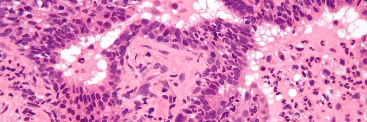
Another interesting stain is the silver stain. Camillo Golgi, a 19th-century Italian physician and Nobel laureate, developed silver staining to study the nervous system. (The cellular organelles known as Golgi bodies are named after him.) There are two classes of cells that take up silver: the argentaffins and the argyrophiles.

Argentaffin
Origin: Latin, argentum (silver) + affinis (akin/affinity)
Affinity for silver; more specifically, cells that take up silver without a reducing agent

Argyrophile
Origin: Greek, argyros (silver) + philia (love)
Love of silver; more specifically, cells that take up silver and need a reducing agent

There are various methods for preparing different silver stains. These stains can be used for a number of applications such as:

  1. Diagnostic microbiology (staining of fungi such as Pneumocystis jiroveci)
  2. Neuropathology (staining of neuritic plaques and neurofibrillary tangles in Alzheimer disease)
  3. Histology (staining of type III collagen and reticulin)

silver stains

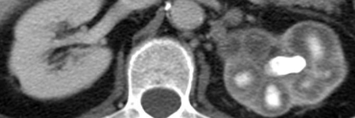
Left: neurofibrillary tangles of Alzheimer disease. Right: membranous nephropathy

The last stain I will briefly touch upon is the periodic acid-Schiff (PAS) stain. This stains for polysaccharides, glycoproteins, glycolipids, and the like. Hence, it can be used to diagnose things like the glycogen storage diseases, adenocarcinomas (which often secrete mucin), Whipple’s disease, etc. The reason why I bring this up is that many people say it incorrectly. It is not periodic as in “recurring time intervals.” Rather, is per-iodic, i.e., of the periodiate ion (IO4)! Periodic acid oxidizes vicinal [L., vicinus (near)] diols to create a pair of aldehydes. These aldehydes then react with the Schiff reagent to produce a purple color.

Here is a clinical scenario: a patient comes to your office for a routine examination. While examining the skin, you notice an ill-defined and oddly-colored brown spot on the patient’s back. Or perhaps while palpating the thyroid gland, you palpate something that doesn’t feel quite right. What is the next step in this patient’s care? If you thought to perform a biopsy, you’d be correct. Indeed, to derive insights from what a patient is suffering from, clinicians obtain a sample of tissue via biopsy from the suspect organ for the tissue to be studied histologically.

Biopsy
Origin: Greek, bios (life) + opsia (a sight)
A sight of life

Histology
Origin: Greek, histos (web/tissue) + logos
The study of tissue

This tissue can initially be examined grossly [L., grossus (thick/coarse)], i.e., by the naked eye, but clinicians often require insights from a pathologist to more completely characterize and understand the tissue they obtained. What does a pathologist study? Let’s take a look:

Pathology
Origin: Greek, pathos (suffering) + logos (word, taken to mean “study of”)
The study of suffering

This is a very generalized definition of pathology (which is absolutely wonderful to me), but let me offer my own comprehensive/digestible definition of pathology. Pathology is the molecular, histological, and gross study of disease processes and their clinical manifestations. Thus, staying true to its etymology, pathology is the systematic and scientific study of suffering. There are four main areas that pathologists explore to better understand disease: aetiology, pathogenesis, morphological changes, and clinical manifestations.

spitz papillary

Left: Spitz nevus (mole). Right: papillary thryoid carcinoma

Aetiology
Origin: Greek, aitos (cause) + logos
The study of the cause of a disease

Pathogenesis
Origin: Greek, pathos + genesis (origin/source)
The origin and development of disease

If all goes well, the pathologist will generate a diagnosis and a prognosis regarding the patient’s condition, and the clinician or the surgeon will proceed accordingly.

Diagnosis
Origin: Greek, dia– (through) + gignoskein (to know) + –osis (condition of)
A condition of knowing through, i.e., a distinguishing

Prognosis
Origin: Greek, pro- (for/before) + gignoskein + –osis
A condition of foreknowledge, i.e., the likely outcome

What tools does a pathologist use to actually study the tissue in question and generate a diagnosis? You’ll have to stay tuned to find out!

The human nervous system is a marvelously complex construct. Through the ages, we have discovered much about the natural world and how to manipulate it, but the human brain and its subsidiaries have not been thoroughly understood despite great efforts. In a previous post, I presented the layers of the brain’s protective coverings—the dura, arachnoid, and pia—and now I would like to take a look at the brain itself. The brain is divided into four lobes: frontal, parietal, temporal, and occipital.

Frontal
Origin: Latin, frons (brow/front)
Belonging at the front, i.e., the brow/forehead

Parietal
Origin: Latin, paries (wall)
Belonging to the walls; in this case, the wall is the top of the skull

brain lobes rev

Temporal
Origin: Latin, tempora (temple of the head)
Belonging to the temples, i.e., the part of the head between the forehead and the ear

Occipital
Origin: Latin, oc– (variant of ob, for towards/against/back of) + caput (head)
Belonging to the back of the head

There are two main branches of the nervous system:

  1. Central nervous system, which consists of the brain and the spinal cord
  2. Peripheral nervous system, which consists of everything else

The building blocks of both nervous systems are neurons, which are colloquially known as brain cells. There are two general classes of neurons: sensory and motor. A sensory neuron—quite logically—allows for the recognition of one of the five senses, e.g., the neurons in the optic nerve receive optical sensory input. A motor neuron delivers the impulse to a muscle spindle to allow for movement, e.g., flexing the biceps. Where does the word neuron come from?

Neuron
Origin: Greek, neuron (sinew/nerve)
So named because nerves have the appearance of sinew

And it turns out that nerve also means sinew, but comes from Latin. This is likely from the observation that a nerve (upon exposure through dissection) looks like sinew! What about the parts of a neuron? A neuron consists of its dendrites, which receive information from other neurons. As for the neuron cell body itself, it is called the soma or the perikaryon. Let’s run through these guys.

Dendrite
Origin: Greek, dendron (tree)
So named because the dendritic processes collectively resemble a tree (see below image)

neuron rev

Soma
Origin: Greek, soma (body)
Refers to the cell body

Perikaryon (plural perikarya)
Origin: Greek, peri– (around) + karyon (kernel, taken to mean nucleus)
Around the kernel/nucleus, i.e., the cell body

I’m deliberately leaving out the axon, myelin, nodes of Ranvier, and Schwann cells because there is more to the story, so stick around!

At some point in our lives, we inevitably fall into some form of ill health. This usually necessitates a visit to a local physician or the hospital, and it may sometimes necessitate a visit from emergency medical services. Thus, it is not unreasonable to assume that most of us have seen the symbol of medicine in one of these settings as a logo of sorts. Except there is a slight problem: there are two snakes on that staff. That staff is, in fact, not the symbol of medicine. It is instead the caduceus, the staff of Hermes. The caduceus was the symbol of commerce, for Hermes is the god of commerce (and thieves…). The U.S. military adopted the caduceus as its symbol of medicine in the 1850s, and it erroneously stuck. The real symbol of medicine is the rod of Ascelpius (Gk. Asklepios).

rod vs caduceusAsclepius was a son of Apollo, and Asclepius took on Apollo’s role as the god of healing in the Greco-Roman pantheon. The etymology behind his name is contested, so I unfortunately have no insights on that matter. There are multiple versions of the story regarding how Asclepius acquired his legendary abilities, and one of the more common ones is this: Asclepius was once asked to cure a man named Glaucus. While doing so, a snake slithered up his staff, and Asclepius promptly killed it. Another snake slithered by with an herb in its mouth and Asclepius placed the herb in the dead snake’s mouth. Sure enough, the dead snake returned to life! Regardless of his “origin story,” long story short, he learned how to raise the dead. Using a snake as a symbol of healing is somewhat paradoxical, since in those days, a snake bite was a death sentence. But Asclepius, utilizing his newfound knowledge, could cure even snake bites—and was thus eventually deified as the god of medicine.

asclepius vs zeus

Asclepius (left) and Zeus (right) share many facial features in classical sculptures

Asclepius was so skilled in medicine that he was asked by Artemis to revive Hippolytus, the son of Theseus and Hippolyta. He was able to do so successfully, and when Hades got word of this, he complained to Zeus that Asclepius was robbing him of his souls. Zeus struck Asclepius down with a thunderbolt to end this traveshamockery (travesty + sham + mockery). Apollo was so enraged that he killed the Cyclopes (singular: Cyclops), who fashioned Zeus’s thunderbolts. Outraged in turn, Zeus forced Apollo to be a servant to King Admetus of Thessay for one year. When Apollo returned to Olympus, Zeus revived the Cyclopes and resurrected Asclepius, turning him into a god.

Outside of the myth, why a snake coiled around a stick? One theory is that the symbol represents the painful process of treating dracunculiasis, better known as Guinea worm disease.

Dracunculiasis
Origin: Neo-Latin, draco (dragon) + -cule (diminutive) + -iasis (condition of)
Condition of small dragons/serpents

Perhaps a picture would illustrate why this makes sense:

guinea worms rev

As a side note, former president Jimmy Carter has been monumental in the global effort to eradicate the Guinea worm, for dracunculiasis is a truly wretched condition. He was recently interviewed and said “I would like the last Guinea worm to die before I do.” (Jimmy Carter was recently diagnosed with melanoma with metastases to the brain and liver.)

Back to our story: many temples were built throughout Greece dedicated to Asclepius. Those who were sick would come and spend a night at the Asclepion, and Asclepius would visit the patient in his or her dreams and relay to them the cure to their disease. One other interesting tidbit is that there were hordes of nonvenomous snakes, colloquially called Aesculapian snakes (Zamenis longissima), that would slither around the temples to promote healing!

Meningitis (plural meningitides) is a nasty condition. From the first post, we already know that meningitis is defined as the inflammation of the meninges. The meninges (singular meninx) are the protective coverings of the brain, and there are three layers, listed outermost to innermost:

  1. Dura mater (Latin, “hard mother,” since it is the thickest layer)
  2. Arachnoid mater (Neo-Latin, “spider-like mother,” since it resembles a spider web)
  3. Pia mater (Latin, “pious mother,” erroneous translation from Arabic umm raqiqah for “tender mother”)

The dura mater is also known as the pachymeninx and the arachnoid mater + pia mater are also known as the leptomeninges.

meninges

Pachymeninx
Origin: Greek, pachy (thick) + meninx (membrane)
Thick membrane, i.e., the dura mater

Leptomeninges
Origin: Greek, leptos (thin) + meninges (membranes)
The thin membranes, i.e., the arachnoid mater and the pia mater

Meningitis can be caused by bacteria, viruses, autoimmune phenomena, or trauma. If the inflammation is caused by bacteria, it is said to be (quite logically) a bacterial meningitis, but if it is caused by the latter three, it is said to be an aseptic meningitis.

Aseptic
Origin: Greek, a- (without) + sepein (to cause decay)

Though the other aetiologies “cause decay,” septic is reserved for bacteria in the medical lexicon. It is important to note that the aseptic meningitides, though wretched, do not typically cause mortality. The bacterial meningitides, however, must be identified and treated swiftly, for they have a relatively high mortality. It is for this reason that if a case of bacterial meningitis is suspected, antibiotics are indicated for immediate use, whether or not the patient truly has a bacterial meningitis. Neutrophils in the cerebrospinal fluid (CSF) is a classic diagnostic marker of bacterial meningitis:

csf pleo rev

There are numerous signs that can hint towards a patient suffering from meningitis, but two of the classic ones are nuchal rigidity (neck stiffness) and photophobia.

Nuchal
Origin: Arabic, nukah (spine)
Used in English as an adjective for the neck

Photophobia
Origin: Greek, photos (light) + phobos (fear) + -ia (condition of)
Condition of the fear of light

When you think “vessel” and “neck,” what words come to mind? For me, the first two words I think of are carotid and jugular. I’ve heard these words used in the vernacular every so often, especially the latter. If someone wants to convey a sentiment of “going for the kill” or delivering a coup de grace, that person can say he or she is “going for the jugular.” Let’s start with this one:

Jugular
Origin: Latin, jugulum (throat)
Named for the fact the jugular veins are near the throat

jugular

There are two jugular veins, one external and one internal on each side of the body. The internal jugular is covered by the sternocleidomastodeus muscle, while the external one is readily visible. These two veins are extremely important because they drain the deoxygenated blood from the head back to the superior vena cava.

What about the other vessel(s) of interest? There is a common carotid artery on both sides of the body, and this artery branches into the internal and external carotid arteries. The former supplies blood to the brain, while the latter supplies blood to the face and head. The etymology is rather interesting:

Carotid
Origin: Greek, karos (stupor)
So called from Galen’s observation that their compression causes stupor/somnolence

carotid rev

If one were to bilaterally massage both carotid sinuses (the point at which the common carotid bifurcates), the baroreceptors of the sinus are fooled into thinking there is a higher-than-normal blood pressure. To compensate, the blood pressure decreases, resulting in a sensation of stupor. Interestingly enough, there is a therapeutic use for this maneuver: the alleviation of supraventricular tachycardia:

Supraventricular tachycardia
Origin: Latin, supra– (above) + ventricle (chamber of the heart; translates to belly, for the ventricles comprise the “belly” of the heart)
Origin: Greek, tachys (swift) + kardia (heart)
A fast pulse due to improper electrical activity above the level of the ventricles

What other afflictions could these vessels suffer from? One thing that can go wrong with the carotid artery is carotid artery stenosis:

Stenosis
Origin: Greek, stenos (narrow/close) + -osis
Condition of narrowness, taken to mean a narrowing

Carotid artery stenosis is oftentimes caused by atherosclerosis, or the reduction of the interior arterial (luminal) diameter due to the accumulation of gunk. This gunk is a lovely mixture of dead cells, cholesterol, and fats.

Atherosclerosis
Origin: Greek, athres (gruel) + sklerein (to harden) + –osis
Condition of the hardening of gruel

Sounds delicious!

What on earth is pyelonephritis? Let’s understand what it actually is by taking a look at its etymological breakdown.

Pyelonephritis
Origin: Greek, pyelos (basin, taken to mean pelvis) + nephros (kidney) + –itis (inflammation of)
Inflammation of the basin and the kidney

At first, this seems cryptic. What does basin refer to, and what exactly does kidney refer to? Starting with the root pyelo-, it denotes “pelvis,” but its base etymology comes from the Greek pyelos (basin). There is in fact such a thing as pyelitis, which would be inflammation localized to the renal pelvis. As for the nephr- root, it of course means kidney, but to be more precise, it refers to the kidney parenchyma. Nephritis on its own denotes the inflammation of the kidney’s functional tissue.

kidney anatomy rev

At the end of the day, pyelonephritis is the joint inflammation of the renal pelvis and the renal parenchyma. A vast majority of the cases are caused by a bacterial agent such as E. coli., and as such, they proliferate in a large portion of the kidney instead of being localized to only the pelvis or the parenchyma.

There are three types of pyelonephritis: acute, chronic, and xanthogranulomatous. Let’s look at the last one:

Xanthogranulomatous
Origin [1]: Greek, xanthos (yellow)
Origin [2]: Latin, granum (grain) + –ula (diminutive) + –oma (mass/proliferation/tumor)
Forming yellow granulomas

Indeed, under the microscope, one would find granulomas—collections of macrophages that try to wall off infections they cannot eradicate. On gross appearance, these granulomas appear yellow because they are macrophages that have been filled to the brim with lipids.

xantho rev

When learning organic chemistry, one of the first topics presented is the naming of organic compounds. The International Union of Pure and Applied Chemistry (IUPAC) codified its recommendations in A Guide to IUPAC Nomenclature of Organic Compounds in 1900. Since then, it has been constantly revising its naming schema for both organic and inorganic compounds. Students of organic chemistry also learn the original nomenclature (“common” nomenclature) for some compounds and groups. First of all, what is the etymology of nomenclature?

Nomenclature
Origin: Latin, nomen (name) + culator (one who calls; from calare, to call)
A calling by name

The IUPAC, for one- and two-carbon alkyl substituents, kept the original nomenclature. For the three- and four-carbon alkyl substituents, they modified them slightly. The rest of the substituents have new standard names, but in many instances, especially in biochemistry, the original names are kept. A few of these names beg for an etymological breakdown.

-CH3; methyl
Origin: Greek, methu (wine) + hule (wood)
Wood spirits/wood alcohol; methanol is a byproduct when destructively distilling wood

-C2H5; ethyl
Origin: Latin, aether (upper air) + -yl (shorter form of hule)
Back-named from (diethyl) ether, for its very low boiling point

-C3H7; propyl
Origin: Greek, pro– (first) + pion (fat) + –yl
First fat, from Dumas’s observation that propionic/propanoic acid is smallest carboxylic acid to display the properties of other fatty acids

linoleic acid revBonus: linoleic [Origin: Greek, linon (flax) + oleum (oil)]

-C4H9; butyl
Origin: Latin, butyrum (butter) + –yl
So named from the fact that rancid butter contains butyric/butanoic acid

-C5H11; pentyl
Origin: Latin, penta– (five) + -yl
Five carbons in the alkyl group

And starting with pentyl, the rest of the alkyl groups are named by Latin numbers. Sad…

Diabetes is a household name in this day and age. More often than not, people are concerned about their sugar intake in order to prevent diabetes. A quick Google Scholar search will reveal that a high sugar intake does not increase the risk of developing diabetes. But, of course, a chronically-high concentration of glucose in the blood will lead to a variety of problems down the line. An age-old clinical observation of diabetes is the tendency to urinate often, termed polyuria [Greek: poly (many) + ourein (to urinate)]. This is in fact where the word diabetes comes from:

Diabetes
Origin: Greek, dia- (through) + banein (to pass)
To pass through, siphon

What about the two words appended to the end of “diabetes”? They are the following:

Mellitus
Origin: Latin, mel (sweet thing)
Something sweet, honey

Insipidus
Origin: Latin, in- (without) + sapere (to have taste)
Lacking taste, in this case sweetness

Diabetes mellitus (DM) has two types, Type 1 and Type 2. The former is usually ascribed to insulin insufficiency, whereas the latter is usually ascribed to insulin resistance. It is given the name “mellitus” because of glucose in the urine, or glucosuria. The large amounts of glucose in the blood finds its way to the urine, and as a result, makes the urine—the entity being passed through—sweet. The polyuria is caused by the osmotic effect of water “following” the glucose. The quick and easy way that physicians would diagnose diabetes mellitus was to simply taste the urine of a patient suspected to have this condition.

vasopressin rev

On the contrary, diabetes insipidus (DI) is caused by a lack of water control and is not related to diabetes mellitus. This “lack of water control” involves problems with anti-diuretic hormone (ADH) production or ADH insensitivity. The former is termed central DI, since it originates in the central nervous system, and the latter is termed nephrogenic [Greek: nephros (kidney) + -genes (born from)] DI. This lack of mechanistic control stimulates profound thirst to replace the massive amounts of water lost in the urine since the water is not being reclaimed by the distal convoluted tubule and collecting duct of the nephron. However, blood glucose is not affected by this condition, there is no glucosuria, and in turn there is no sweetness in the urinehence the urine is without taste, or “insipid.”

In colloquial language, we often append the suffix ­-itis to a word to denote a disease or condition of said word. It is in effect an indication of an unhealthy amount of something either abstract or tangible. If someone is being lazy, we can say that he or she has come down with a case of lazyitis. Too much time in front of a computer can afflict you with a nasty case of computeritis. At its roots, –itis means “pertaining to” in Greek, so in some sense, this colloquial use of the suffix is the most faithful to its base etymology.

In more technical language, –itis is used as a medical suffix, and it simply denotes “inflammation of.” I can’t tell you how many times I have heard people use ­–itis to immediately denote “infection of.” Let’s list some examples.

Appendicitis
Origin: Neo-Latin, appendix (something appended, in this case to the colon) + -itis (inflammation of)
Inflammation of the appendix

Bursitis
Origin: Greek, byrsa (hide/skin) + ­-itis
Inflammation of the bursa (a pouch or sack that facilitates motion, e.g., suprapatellar bursa)

Cholecystitis
Origin: Greek, chole (bile) + kystis (bag) + -itis
Inflammation of the gall bladder (which stores bile)

Yes, many times, these conditions come about from infection by a bug that isn’t supposed to be there. But there are many other causes of inflammation, such as:

  1. Trauma
  2. Autoimmune reaction
  3. Drugs
  4. Burns
  5. Radiation

So the next time someone mentions an ­–itis, always remember that the inflammation of the target organ can be caused by something other than infection. If you wish to convey the fact that an inflammation of the appendix is caused by an infection, you can prepend the word with “infectious,” to make it an infectious appendicitis. Or if you are more certain of the cause of this infection, you can use a bacterial/viral/fungal modifier, for a bacterial appendicitis. For a non-infectious cause, you could say the cause for an inflammation of the liver is a drug-induced hepatitis.

Finally, if someone mentions a type of ­­itis that you are not familiar with, use the word itself to help you out. I remember hearing the term bronchiolitis last year when learning about COPD (chronic obstructive pulmonary disease). I didn’t know what bronchiolitis was until I stopped for a second to think about it: the inflammation of the bronchiole(s). Contrast this with bronchitis, which is the inflammation of a main stem bronchus.

lung anatomy rev

So, given this, let me know in the comments what these terms are and what their most common causes are:

  1. Cholangitis
  2. Colitis
  3. Pneumonitis
  4. Endocarditis
  5. Vasculitis
  6. Phlebitis
  7. Gastroenteritis